Advice after your nerve block for surgery
On this page…
Introduction
We have been advised by your hospital consultant that you need to have a Renal cryoablation for the treatment of a tumour in a kidney. These are done in the radiology department by an Interventional Radiologist (doctor specialising in medical imaging) by guiding a needle into the body under CT control. The procedures will be performed by a radiologist who will be assisted by radiographers and radiology nurses.
This information tells you about having the procedure, what is involved, and what the possible risks are. This leaflet may not answer all your questions and it is not meant to replace informed discussion between you and your doctor, but can act as a starting point for such discussions. If you have any questions about the procedure, please ask the doctor who has referred you or the department that will be performing it.
What is a Renal Cryoablation?
Other tests that you have had will have shown that you have a tumour in your kidney. Your urologist or oncologist will have discussed this with you, and in your case, you and the medical team (the urologist and an interventional radiologist who is going to perform the procedure) have decided that cryoablation is the best treatment option.
Cryoablation is a technique that destroys tissue, in this case through freezing. In order to produce the freeze, needles are placed into the kidney using CT guidance. A mixture of gases are then used to freeze and thaw the tips of the needles. Temperatures lower than - 100°Celsius are produced, but this only travels a few centimetres so most of the normal kidney tissue is not affected. If necessary, the procedure can be repeated.
What is a CT scan?
A CT scan is an examination using X-rays by lying on a flat table inside a scanner that produces detailed images of your body. These images are cross-sectional (like slices) and are used to produce 2D and 3D images of the body’s organs.
Before your appointment
Please let us know if:
- you are taking any medication which impairs the bloods ability to clot (anticoagulation or blood thinning drugs) e.g. Warfarin or clopidigrel, or, if you are aware that you may have bleeding problems. The radiologist who will be performing your examination can be notified and may need to discuss this with you and your hospital consultant before your appointment can be confirmed as the medication may need to be stopped or changed before the procedure
- there is any possibility that you may be pregnant. Please be aware, that all patients from 12-55 years of age (regardless of gender) who will undergo a Radiology procedure will be asked about the possibility of them being pregnant.
- you are diabetic - please note that you are required to starve before the examination. You may be given an early morning appointment or alternative medication will be given on the ward.
Eating and Drinking
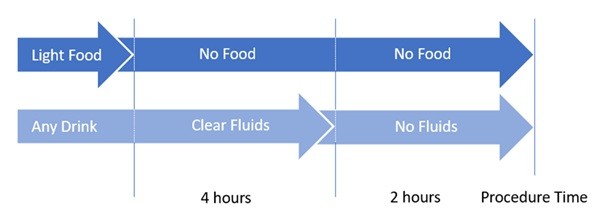
You should have nothing to eat 6 hours prior to the time of your procedure. You can continue to drink clear fluids (water) up to 2 hours prior to the procedure but then nothing should be consumed.
Special Needs
If you have any special needs that we need to consider, such as:
- requiring the Vascular Access Team for injections
- requiring Hospital transport
- having limited mobility and / or requiring a hoist
- you will need an interpreter. We are not able to use family members or friends to interpret.
In order to make your appointment run more smoothly, please let us know if any of these apply, in advance, by calling our bookings team (contact details are displayed on the main Radiology page).
What to bring with you
You will usually be admitted for the procedure as a day case. Occasionally it is necessary for patients to stay overnight in hospital after the biopsy. For this reason we advise that you prepare an overnight bag and bring this with you. You may wish to bring your own dressing gown, slippers and reading material.
Please bring with you to your appointment any sprays or inhalers that you are taking. Please bring a list of any regular medications you are taking
During the procedure
Referral and consent
You will be asked to sign a consent form confirming that you understand the procedure and are aware of the risks and benefits involved. Where practical this is usually done on the ward, before you come to the Radiology Department or in an outpatient clinic environment before your admission to hospital for the procedure.
The procedures are normally done as a planned procedure and you should have plenty of time to discuss the situation with your doctor or the nurse specialist looking after you before you sign the consent form. When you arrive for the procedure you will also discuss the procedure with the radiologist who will be performing the procedure who will again sign the consent form with you.
If after discussion with your hospital doctor or radiologist, you do not want the procedure carried out, then you can decide against it. If the radiologist feels that your condition has changed or that your symptoms do not indicate such a procedure is necessary then they will explain this to you and they will communicate with the referring doctor who will review your situation as soon as possible. At all times the radiologist and referring doctor will be acting in your best interests.
Procedure
You will need to wear a hospital gown. The Porters will collect you from the ward and bring you to the CT Department, on your bed, for the procedure.
The procedure will be explained to you by the radiologist. You will be able to ask any further questions that you may have. The anaesthetist will also see you before the procedure and talk to you about the sedation or general anaesthetic. If you have any questions or concerns, this is the time to ask
Once the anaesthetist has put you to sleep you will be moved into the CT scanner, positioned lying on your front and be monitored throughout the procedure. The radiologist will keep everything as sterile as possible and will wear a theatre gown and operating gloves. The skin near the point of needle insertion will be thoroughly cleaned with antiseptic, and then most of the rest of your body covered with a theatre towel.
Using CT the needles will be guided into the kidney and tumour. The tumour will then be frozen to destroy it. A completion scan is then performed to assess the immediate results of the treatment. Generally, cryoablation itself will take around 2-3 hours, but on occasion it may take longer. This is variable depending on the complexity and size of the tumour.
After the procedure
When you wake up from your general anaesthetic, you will be in the recovery area. The nurse will regularly check your pulse rate and blood pressure. Once you are comfortable and your blood pressure is stable, you will be taken to the ward for an overnight stay. On the ward, you will gradually be allowed to drink water. If you are able to tolerate good amounts and do not feel sick, then you will be able to have a hot drink and something light to eat. You will have an intravenous drip in your arm, which will be removed before you go home. Your nurse will offer you pain relief to help with any discomfort.
Normally, you will able to go home the same day as your procedure. Before you go home we will discuss your follow-up treatment with you. You should expect to be off work for one week after the treatment. You will have an appointment to come back into clinic after the procedure
Risks
As with any procedure or operation, complications are possible. The possibility of these occurring will vary for each patient and the possibility of these happening to you will be discussed with you, before you sign the consent form.
Bleeding: Any intervention on a blood vessel carries a risk of bleeding. There may be a small bruise around the site where the needles have been inserted and this is quite normal. There is a chance that the bruise may become very large and uncomfortable, but this does not happen very often.
Pain: This can occur due to the necrosis of the tumour due to the ablation. This is usually worst in the first 12 hours. This can be controlled by pain killers and you may be given further tablets to take home with you.
Temperatures: Most patients get a slight fever after the procedure. This is a good sign as it means that the tumour is breaking down. The pain killers you will be given will help control this fever.
Blood in the urine: A few patients get blood in the urine after the procedure and this can persists for approximately one week. However, if the urine becomes offensive (strong smell or colour) and if it is associated with a high fever and feeling unwell, there is the possibility of infection and you should contact your urologist
Radiation: X-rays used in the procedure are a type of radiation. We are all exposed to natural background radiation every day from the sun, food we eat, and the ground. Exposure to medical X-rays carries a small additional risk, but your doctor feels that this risk is outweighed by the benefits of having the procedure and the amount of radiation you will receive is kept to as low as reasonably possible
Contact us
If you have any queries relating to this information, please contact the Radiology service.
About this information
Service:
Radiology
Reference:
PILS-CT.12
Approval date:
2 October 2024
Review date:
2 October 2027
Click ‘show accessibility tools’ at the bottom of the page

Then click ‘select language’
![]()
Alternative formats
You can use the accessibility toolbar at the bottom of your screen to:
-
Change the text size
-
Adjust the font
-
Modify the colour contrast
-
Use the translate function
If you would like this information in another format, such as Braille, audio, or easy read, please speak to a member of staff.
You can also print as well as download as PDF using the “Print this page” button at the end of the page.
Staff will print a copy for you on request
Important note
This page provides general information only. It is developed by clinical staff and is reviewed regularly every 3 years for accuracy. For personal advice about your health, or if you have any concerns, please speak to your doctor.