Advice after your nerve block for surgery
On this page…
Intervention Radiology – Biliary Drainage Procedure
We have been advised by your hospital consultant that you need to have a biliary drainage procedure performed in the Radiology (X-ray) Department under ultrasound and fluoroscopic control.
These are done in the radiology department by an Interventional Radiologist (doctor specialising in medical imaging), by guiding a needle into the body under ultrasound and fluoroscopic control, to access a particular organ to be able to insert drains, stents or to obtain more detailed images.
This information tells you about having the procedure, what is involved, and what the possible risks are. This is not intended to replace the discussion between you and your consultant, but may act as a starting point for discussion.
If after reading this information you still have concerns or require further explanation, please contact us using the telephone number on your appointment letter, or ask the ward staff.
What is the procedure?
One of the normal functions of the liver is to produce bile which is important for digestion. The bile drains from the liver to the bowel through a series of small ducts into the gastrointestinal tract.
If the bile duct becomes blocked, then bile cannot drain normally and jaundice (yellowing of the skin) may develop. To treat the jaundice, the bile needs to be drained from the liver by inserting a drain to externally drain out the bile or placing a stent through the blockage to open it up.
A Percutaneous Transhepatic Cholangiogram (PTC) is the process of taking pictures of the bile ducts to see where the blockage might be.
Alternatively, the gallbladder may be inflamed (cholecystitis) due to an obstruction of its drainage, and a drain can be inserted into the gallbladder to drain it, (Cholecystostomy), or images of an existing drain (cholecystostogram) may be required.
The procedure will be performed by a radiologist (doctor specialising in medical imaging) who will be assisted by radiographers and radiology nurses.
What is Fluoroscopy?
Fluoroscopy is a process that uses X-rays to produce a real time moving image on a television monitor.
What is Ultrasound?
An ultrasound scan is an examination that uses sound waves to take pictures of the part of the body being examined. It does not involve X-rays.
Before your appointment
Please let us know if:
- you are taking any medication which impairs the bloods ability to clot (anticoagulation or blood thinning drugs) e.g. Warfarin or clopidogrel, or, if you are aware that you may have bleeding problems. The radiologist who will be performing your examination can be notified, and may need to discuss this with you and your hospital consultant before your appointment can be confirmed, as the medication may need to be stopped or changed before the procedure. The Interventional nursing team can be informed on 0300 613 2161 (Frimley) or 0300 615 3544 (Wexham).
Please also let us know if:
- you have limited mobility and will require assistance to get on and off the scanner.
- you will need an interpreter. We are not able to use family members or friends to interpret.
- there is any possibility that you may be pregnant. All patients from 12-55 years of age (regardless of gender) who will undergo a Radiology procedure will be asked about the possibility of them being pregnant.
In order to make your appointment run more smoothly, please let us know if any of these apply, in advance, by calling our bookings team (contact details are displayed on the main Radiology page).
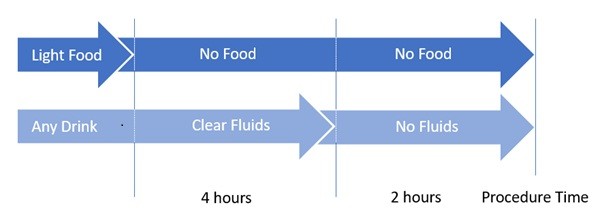
Eating and Drinking
You should have nothing to eat 6 hours prior to the time of your procedure. You can continue to drink clear fluids (water) up to 2 hours prior to the procedure but then nothing should be consumed.
Special Needs
If you have any special needs that we need to consider, such as:
- requiring the Vascular Access Team for injections
- requiring Hospital transport
- having limited mobility and / or requiring a hoist
In order to make your appointment run more smoothly, please let us know if any of these apply, in advance, by calling 0300 613 4140 between 9:00am and 4:00pm, Monday to Friday.
If a BSL interpreter has been booked for this appointment and you require any further information, please contact the team on 07918 375794 (this number will accept a text message).
What to bring with you
You will usually be admitted for the procedure as a day case. Occasionally it is necessary for patients to stay overnight in hospital after the procedure. For this reason we advise that you prepare an overnight bag and bring this with you. You may wish to bring your own dressing gown, slippers and reading material.
Please bring with you to your appointment any sprays or inhalers that you are taking. Please bring a list of any regular medications you are taking
During the procedure
Referral and consent
You will be asked to sign a consent form confirming that you understand the procedure and are aware of the risks and benefits involved. Where practical this is usually done on the ward, before you come to the Radiology Department or in an outpatient clinic environment before your admission to hospital for the procedure.
The procedures are normally done as a planned procedure and you should have plenty of time to discuss the situation with your doctor or the nurse specialist looking after you before you sign the consent form. When you arrive for the procedure you will also discuss the procedure with the radiologist who will be performing the procedure who will again sign the consent form with you.
If after discussion with your hospital doctor or radiologist, you do not want the procedure carried out, then you can decide against it. If the radiologist feels that your condition has changed or that your symptoms do not indicate such a procedure is necessary then they will explain this to you and they will communicate with the referring doctor who will review your situation as soon as possible. At all times the radiologist and referring doctor will be acting in your best interests.
Procedure
You will need a cannula (small tube) inserted into a vein in your arm to allow access for fluids and for administering medication.
You will need to wear a hospital gown. The Porters will collect you from the ward and bring you to the X-ray Department, on your bed, for the procedure.
The procedure will be explained to you by the radiologist. You will be able to ask any further questions that you may have.
You will be asked to lie on the examination table. It is important that you stay very still until the procedure is over. If you are uncomfortable please let the doctor know.
The ultrasound will be used to decide on the most suitable place for inserting the needle.
Everything will be kept sterile and the radiologist will wear sterile gloves. Your skin will be cleaned with antiseptic solution and sterile drapes will be placed over this area. Then your skin will be numbed with local anaesthetic. When the local anaesthetic is injected, it will sting to start with, but this soon wears off, and the skin and deeper tissues should then feel numb.
The radiologist will insert the thin needle whilst looking at the images to ensure accuracy of positioning. You may be aware of a pushing sensation as the needle is positioned, but this is generally done so quickly that it does not cause much discomfort. You may be asked to hold your breath to help the needle to be positioned accurately.
A needle followed by a wire and catheter (fine plastic tube) will be inserted into the bile ducts using ultrasound and x-ray guidance. A small amount of x-ray dye (contrast agent) is injected to allow images to be taken of the ducts. The procedure may finish at this stage or a stent or drainage catheter may be inserted, this will depend on what the radiologist finds.
If the procedure does become painful you should tell the radiologist performing the procedure and they may give you more local anaesthetic or sedatives.
After the procedure
When the procedure is finished, a small dressing may be placed to cover the skin incision or you may have a drain attached to a collecting bag and you will be returned to the ward.
Once you have returned to your ward, nurses will carry out close and regular observations, such as taking your pulse, blood pressure and temperature, to make sure that there are no problems. You may be required to stay in bed for at least four hours. You should tell the nurses if you feel worsening pain or a rise in your body temperature.
You should drink plenty of fluids and eat normally.
If you have an external drainage catheter attached to a bag, it is important that you are aware of it. For example, you should try not to get up out of a chair without remembering about the bag, and making sure that it can move freely with you. However, you will be able to lead a normal life with the catheter in place. The bag needs to be emptied fairly frequently so that it does not become too heavy.
Taking the external catheter out does not hurt and all that remains is a small puncture site which is covered by a small plaster. This should be kept clean and dry before being removed after 2 days.
If you have any problems after the procedure please speak to the staff on the ward or contact your GP or emergency doctor if problems occur when you have returned home.
How long does it take?
As an approximate guide, expect to be in the Radiology Department for about 45 minutes to one hour. However much of this time is taken up by preparation rather than the procedure itself. You will normally be expected to rest on the ward for at least four hours after the procedure. If you have been admitted specifically for this procedure as a day case patient, you should then be able to go home. In some cases you may be asked to stay overnight.
Risks
As with any procedure or operation, complications are possible. We have included the most common risks and complications in this leaflet. The possibility of these occurring will vary for each patient and the possibility of these happening to you will be discussed with you, before you sign the consent form.
Bleeding: Any interventional procedure carries a small risk of bleeding. On rare occasions, this may become severe. There may be soreness around the injection or drain site and some bruising. On rare occasions, bleeding may be severe and require blood transfusion or another radiological procedure to stop it.
Infection: Occasionally there may be infection in the area surrounding the biopsy site. This can usually be treated with antibiotics. If the bile is infected, although you may be on antibiotics, there is a small risk that infection might be released into your bloodstream, making you unwell for a period
Bile leak: If there is a leak of bile for any reason into the abdomen from this procedure, it may require another radiology inserted drain through the skin as a temporary measure until the fluid has gone.
X-rays are a type of radiation. We are all exposed to natural background radiation every day from the sun, food we eat, and the ground. Exposure to medical X-rays carries a small additional risk, but your doctor feels that this risk is outweighed by the benefits of having the test and the amount of radiation you will receive is kept to as low as reasonably possible.
Contact us
If you have any queries relating to this information, please contact the Radiology service.
About this information
Service:
Radiology
Reference:
PILS-IR.14
Approval date:
2 October 2024
Review date:
2 October 2027
Click ‘show accessibility tools’ at the bottom of the page

Then click ‘select language’
![]()
Alternative formats
You can use the accessibility toolbar at the bottom of your screen to:
-
Change the text size
-
Adjust the font
-
Modify the colour contrast
-
Use the translate function
If you would like this information in another format, such as Braille, audio, or easy read, please speak to a member of staff.
You can also print as well as download as PDF using the “Print this page” button at the end of the page.
Staff will print a copy for you on request
Important note
This page provides general information only. It is developed by clinical staff and is reviewed regularly every 3 years for accuracy. For personal advice about your health, or if you have any concerns, please speak to your doctor.