Advice after your nerve block for surgery
On this page…
Anatomy of the Elbow
The elbow joint is formed between the bones of the upper arm (humerus) and forearm (radius and ulna), as well as the muscles, ligaments and tendons surrounding the joint. It is a hinge joint between the humerus and the radius and ulna which allows the forearm and hand to be moved towards (flexion) and away (extension) from the body.
What is an elbow fracture?
An elbow fracture is when there is a break in one or more of the three bones that make up the elbow joint. It normally happens after an injury, like a blow to the elbow, a fall, or a twist. The elbow may also be sprained, strained or dislocated. Fractures can be displaced (where two ends of the bone separate) or non-displaced (where the bone is broken but stays in place). A fracture can be open (where the bone goes through the skin) or comminuted (where the bone breaks in more than two pieces).
How is an elbow fracture managed?
Management will always aim to restore function and movement of the elbow as best as possible. Conservative management: If the bones are well-aligned, then your elbow may be immobilised in a sling, cast or a splint for a number of weeks (usually up to 6 weeks), to allow the bones to heal in the correct position. If your bones are not aligned, then your fracture will need to be ‘reduced’ to position the joint correctly. If this is successful then you will have a cast on your elbow for up to 6 weeks as mentioned above.
The healing of your fracture will be monitored using x-rays in fracture clinic. This will show how well the fracture is healing and you will then be able to progress with exercises and putting weight through the wrist as advised by the doctors or physiotherapists.
Surgical management
If your fracture is unstable, then you may require an operation called an Open reduction Internal Fixation (ORIF); this means that your fracture will be re-aligned and stabilised using metalwork such as plates and screws. This is to ensure the joint heals in the best position to restore function. Your fracture will also be monitored in Fracture Clinic to assess how it is healing.
Things to avoid
You should not weight bear through your operated arm and no lifting. This is normally for six weeks, but you will be advised by your consultant or physiotherapist after your operation. No driving until advised by your consultant.
Post-operation: You are likely to have swelling in your operated arm. Resting and elevating your arm can help reduce swelling.
Post Operative Exercises
Try to complete 10 repetitions of each exercise, 3 times per day to prevent stiffness throughout your other joints.
|
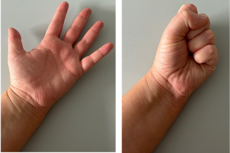
Finger flexion and extension: Bring fingers to hand to make a fist |
|
|
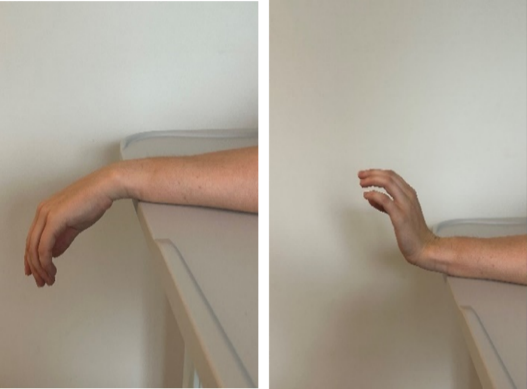
Wrist flexion and extension Bend the wrist backwards and forwards |
|
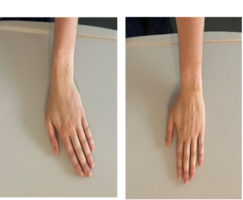
| Wrist radial and ulna denervation: Bend the wrist first towards the little finger, then towards the thumb |
|
|
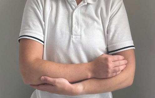
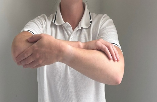
Shoulder pendular: Abduction: In your sling you can cradle the operated arm with your good arm if you need to. |
|
| Flexion: In your sling you can cradle the operated arm with your good arm if you need to. Gently bring the operated arm forwards and back to the start position. |
|
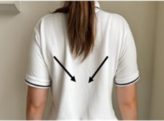
| Scapular setting: Start with your arm relaxed in your sling. Bring the shoulder up and take the shoulder blade slightly back and bring the shoulder blade down. Hold for 5 seconds then relax. |
|
Follow up Physiotherapy
Once you are out of your cast, sling or splint, you may be referred to outpatient physiotherapy from the fracture clinic if you are having trouble getting your elbow moving again. They will work on restoring your range of movement and increasing the strength in your elbow.
Helpful Contacts
Hospital Switchboard – 0300 614 5000
Wexham Park:
- Ward 1 reception – 0300 615 3012
- Ward 2 Orthopaedics – 0300 615 3801
Frimley Park:
- Orthopaedic Helpline – 0300 6132638
- Ward F4 reception – 0300 613 4865
- Ward F5 reception – 0300 613 6698
- Ward F6 reception – 0300 613 4173
Contact us
If you have any queries relating to this information, please contact the Orthopaedics service.
About this information
Service:
Orthopaedics
Reference:
BB/109
Approval date:
1 December 2025
Review date:
1 December 2028
Click ‘show accessibility tools’ at the bottom of the page

Then click ‘select language’
![]()
Alternative formats
You can use the accessibility toolbar at the bottom of your screen to:
-
Change the text size
-
Adjust the font
-
Modify the colour contrast
-
Use the translate function
If you would like this information in another format, such as Braille, audio, or easy read, please speak to a member of staff.
You can also print as well as download as PDF using the “Print this page” button at the end of the page.
Staff will print a copy for you on request
Important note
This page provides general information only. It is developed by clinical staff and is reviewed regularly every 3 years for accuracy. For personal advice about your health, or if you have any concerns, please speak to your doctor.